Home
Uncategories
Shoulder Anatomy Diagram / Shoulder Muscles Anatomy And Functions Kenhub / The shoulder girdle includes three bones—the scapula, clavicle and humerus.
Shoulder Anatomy Diagram / Shoulder Muscles Anatomy And Functions Kenhub / The shoulder girdle includes three bones—the scapula, clavicle and humerus.
Shoulder Anatomy Diagram / Shoulder Muscles Anatomy And Functions Kenhub / The shoulder girdle includes three bones—the scapula, clavicle and humerus.. It is one of the most mobile joints in the human body, at the cost of joint stability. Typically, one attachment remains stationary and is called the origin and the other attachment moves. Tutorials on the shoulder muscles (e.g rotator cuff muscles: The shoulder joint (glenohumeral joint) is a ball and socket joint between the scapula and the humerus.it is the major joint connecting the upper limb to the trunk. Tendons are cords made of tough tissue, and they work as special connector pieces between bone.
Formerly called tendinitis, this is inflammation or irritation of a tendon that attaches to a bone. 2.2 shoulder muscles and shoulder tendons. Labeled anatomy chart of male triceps and back muscles on white background labeled human anatomy diagram of man's arm, shoulder and upper back muscles in a posterior view on a white background. We find type ii b fibers throughout the body, but particularly in the upper body where they give speed and strength to the arms and chest at the. Tutorials on the shoulder muscles (e.g rotator cuff muscles:
Shoulder Anatomy from marvel-b1-cdn.bc0a.com To learn more about how the shoulder muscles work, review the accompanying lesson called shoulder muscles: Shoulder pain, instability and, in some cases, a feeling of grinding, locking or catching while moving the shoulder. Due to the inherent complexity of the shoulder joint, it is also particularly prone to problems. Human anatomy diagram shoulder anatomy shoulder muscles shoulder muscles and chest. Antique illustration of human body anatomy: Tutorials on the shoulder muscles (e.g rotator cuff muscles: The shoulder joint is not very stable, and it may be easily dislocated as the anatomy is conducive to that and the soft tissues around the joint are. Elbow fractures icons orthopedic impingement body yoga anatomy back shoulder elbow fracture glenoid icons pain shoulder and elbow pain shoulder joint.
Plus, exercises for training them.
Elbow fractures icons orthopedic impingement body yoga anatomy back shoulder elbow fracture glenoid icons pain shoulder and elbow pain shoulder joint. Anatomy • free medical books. Diagram of shoulder muscles and tendons. The following is an overview of the shoulder muscle anatomy. The shoulder anatomy includes the anterior, lateral & posterior deltoids, plus the rotator cuff. The muscles in the shoulder aid in a wide. However, more serious injuries, such as complete rotator cuff tears, may require surgical repair. The primary function of the shoulder girdle is to give strength and range of motion to the arm. The shoulder girdle includes three bones—the scapula, clavicle and humerus. The most common symptoms of a torn shoulder labrum are: Ac joint is a diathrodial joint with a fibrocartilaginous disk. This diagram depicts labeled muscle diagram 1024×1878 with parts and labels. Labeled anatomy chart of male triceps and back muscles on white background labeled human anatomy diagram of man's arm, shoulder and upper back muscles in a posterior view on a white background.
The most flexible joint in the entire human body, our shoulder joint is formed by the union of the humerus, the scapula (or shoulder blade), and the clavicle (or collarbone). Anatomy • free medical books. The shoulder is a complex combination of bones and joints where many muscles act to provide the widest range of motion of any part of the body. Contents hide 1 anatomical terms. Contents hide deltoids anatomy.
Shoulder Anatomy Muscles Anatomy Drawing Diagram from www.anatomynote.com 3d tutorial on the anatomy of the shoulder joint from anatomyzone for more videos, 3d models and notes visit: The shoulder girdle includes three bones—the scapula, clavicle and humerus. Shoulder pain anatomy map / anatomy of neck and shoulders anatomy drawing diagram. Labeled anatomy chart of male triceps and back muscles on white background labeled human anatomy diagram of man's arm, shoulder and upper back muscles in a posterior view on a white background. The most flexible joint in the entire human body, our shoulder joint is formed by the union of the humerus, the scapula (or shoulder blade), and the clavicle (or collarbone). 17 photos of the diagram of shoulder muscles and tendons. This diagram depicts labeled muscle diagram 1024×1878 with parts and labels. Formerly called tendinitis, this is inflammation or irritation of a tendon that attaches to a bone.
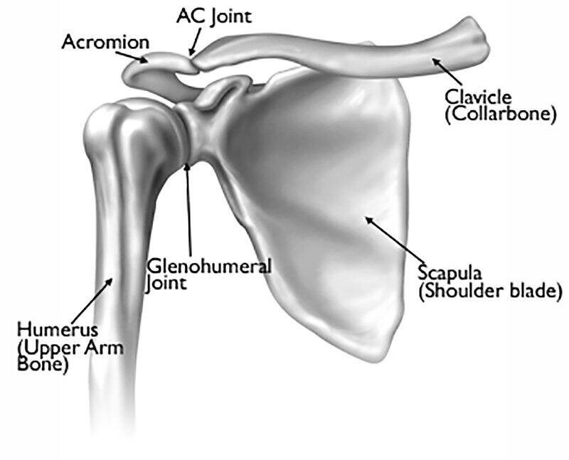
The shoulder joint (glenohumeral joint) is a ball and socket joint between the scapula and the humerus.it is the major joint connecting the upper limb to the trunk.
Diagram of shoulder muscles and tendons. Tendons are cords made of tough tissue, and they work as special connector pieces between bone. Shoulder pain anatomy map / anatomy of neck and shoulders anatomy drawing diagram. Human anatomy diagram shoulder anatomy shoulder muscles shoulder muscles and chest. Formerly called tendinitis, this is inflammation or irritation of a tendon that attaches to a bone. Ac joint is a diathrodial joint with a fibrocartilaginous disk. Learn about these muscles, their origin and insertion points, and their functional anatomy. A second joint in the shoulder is the junction of the collar bone with the shoulder blade, called the. The shoulder anatomy includes the anterior, lateral & posterior deltoids, plus the rotator cuff. The shoulder girdle includes three bones—the scapula, clavicle and humerus. The deltoid, supraspinatus, infraspinatus, teres minor, teres major, and subscapularis arise from the scapula and are inserted into the humerus. Browse 3,854 shoulder anatomy stock photos and images available, or search for shoulder joint or rotator cuff to find more great stock photos and pictures. Numerous muscles help stabilize the three joints of.
Shoulder pain, instability and, in some cases, a feeling of grinding, locking or catching while moving the shoulder. The shoulder joint is not very stable, and it may be easily dislocated as the anatomy is conducive to that and the soft tissues around the joint are. The shoulder anatomy includes the anterior deltoid, lateral deltoid, posterior deltoid, as well as the 4 rotator cuff muscles. The primary function of the shoulder girdle is to give strength and range of motion to the arm. Antique illustration of human body anatomy:
Labeled Anatomy Chart Of Neck And Shoulder Muscles On White Background Stock Photo Download Image Now Istock from media.istockphoto.com The shoulder joint is formed where the humerus upper arm bone fits into the scapula shoulder blade like a ball and socket. Ac joint is a diathrodial joint with a fibrocartilaginous disk. 17 photos of the diagram of shoulder muscles and tendons. Ebraheim's educational animated video describes muscle anatomy of the shoulder girdle and anatomy of the shoulder joint.anatomy of the shoulder muscles a. The shoulder girdle includes three bones—the scapula, clavicle and humerus. Browse 3,854 shoulder anatomy stock photos and images available, or search for shoulder joint or rotator cuff to find more great stock photos and pictures. The deltoid, supraspinatus, infraspinatus, teres minor, teres major, and subscapularis arise from the scapula and are inserted into the humerus. This diagram depicts labeled muscle diagram 1024×1878 with parts and labels.
It is one of the most mobile joints in the human body, at the cost of joint stability.
The shoulder joint is formed where the humerus (upper arm bone) fits into the scapula. Other important bones in the shoulder include: The anterior shoulder pain usually develops when injury or inflammation occurs in the tendons that are attached to the shoulder joint. Elbow fractures icons orthopedic impingement body yoga anatomy back shoulder elbow fracture glenoid icons pain shoulder and elbow pain shoulder joint. Deltoides triangular refers to the front head of the. What does a torn shoulder labrum feel like? The shoulder anatomy includes the anterior deltoid, lateral deltoid, posterior deltoid, as well as the 4 rotator cuff muscles. The following is an overview of the shoulder muscle anatomy. This diagram depicts labeled muscle diagram 1024×1878 with parts and labels. Numerous muscles help stabilize the three joints of. Learn about these muscles, their origin and insertion points, and their functional anatomy. 2.1 bones of the shoulder girdle. 17 photos of the diagram of shoulder muscles and tendons.


0 Comments:
Posting Komentar